Tooth decay in front teeth: How it starts and how AI can help
Learn how front tooth decay starts, its early signs, and treatments. See how AI and computer vision can help dentists detect cavities early.

Tooth decay in front teeth is a common oral health issue that often begins quietly without obvious symptoms. In the early stages, there may be no pain or visible changes, even as enamel starts to weaken beneath the surface.
If left untreated, this slow-moving condition can lead to pain, infection, or even tooth loss. To improve early detection, dentists often rely on medical images such as X-rays, digital scans, and photographs taken inside the mouth. These tools reveal signs that routine visual exams may miss.
However, despite the effectiveness of these tools, human error can still cause early signs of decay to be overlooked. This is where cutting-edge technology is beginning to make a difference.
In particular, computer vision, a branch of artificial intelligence (AI) that focuses on analyzing visual data like medical images, is now making it possible for dentists to interpret scans with greater accuracy.
In this article, we’ll explore what tooth decay is, what causes it, and the various ways it can be treated. We’ll also look at how computer vision is being used to prevent and manage this common dental issue. Let’s get started!
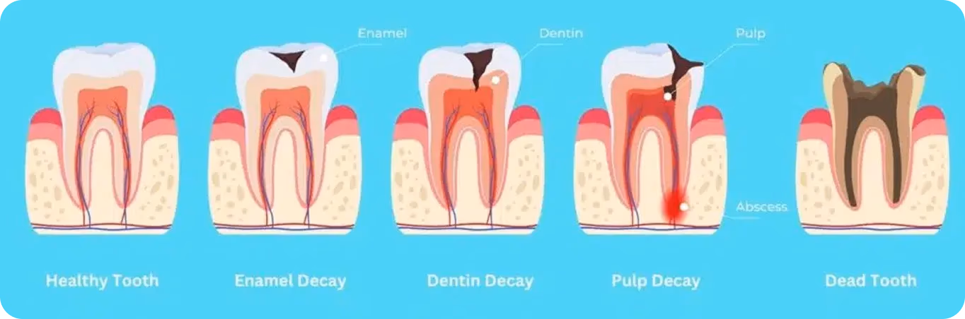
Fig 1. The different stages of tooth decay (Source)
Link to this sectionWhat causes tooth decay in front teeth?#
Cavities in the front teeth usually begin with plaque buildup. Cavities are small holes that form when tooth enamel is damaged by acids produced by bacteria. Plaque forms when food particles and bacteria cling to the surface of the tooth, especially near the gumline. If plaque is not cleared away with daily oral hygiene practices, it can harden into tartar, which raises the risk of decay.
Your diet plays a major role in this process. Sugary foods, sweet snacks, and sugary drinks provide fuel for bacteria. As the bacteria feed, they produce acid that weakens the enamel. Acidic foods such as citrus fruits and sodas make this erosion worse. Once enamel breaks down, small openings form, and decay spreads deeper into the tooth structure.
Saliva usually helps wash away food particles, but people with dry mouths have less natural protection. Poor oral hygiene habits also speed up decay. Skipping floss, avoiding fluoride toothpaste, or neglecting regular dental visits allows damage to progress more quickly. Young children and teens are especially at risk since their permanent teeth are still developing.
Although cavities are more common in back teeth and chewing surfaces, decay in the front teeth needs to be taken care of promptly because these teeth play a key role in oral health and everyday use.
Link to this sectionEarly signs of a front tooth cavity#
The earliest sign of tooth decay in front teeth is often the appearance of white spots on the surface of the tooth. These spots are an early warning that minerals are being lost from the enamel.
As decay progresses, discoloration may follow. Light brown marks can appear and gradually darken, especially on the front surface, where even small changes are noticeable. Because these teeth are always visible, patients often recognize the problem earlier than they would with decay in the back teeth.
Tooth sensitivity is another common symptom. In the early stages, discomfort may occur when eating sweet foods, drinking sugary drinks, or consuming acidic foods. While the sensitivity may begin as mild, it usually worsens as decay moves deeper. Once the dentin or pulp is affected, the pain becomes harder to ignore, and more invasive treatment is required.
Dentists rely on X-rays and regular dental check-ups to confirm these changes. With early detection, patients have a better chance of saving their natural teeth and avoiding advanced procedures.
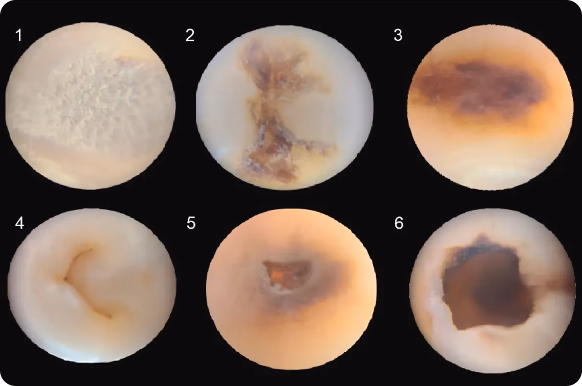
Fig 2. Microphotography of the different stages of tooth decay (Source)
Link to this sectionTreatment options for front tooth decay#
Cavities in the front teeth can be treated in different ways depending on their severity. The care options depend on how far the decay has progressed and the steps needed to protect one’s overall oral health.
Here are some common treatment options that dental professionals recommend:
- Composite fillings vs. tooth-colored fillings: Composite fillings are often used for small cavities because they blend with the natural tooth. Tooth-colored fillings are especially popular for front teeth since they look seamless on visible surfaces.
- Dental bonding, composite resin, and veneers: Dental bonding uses a tooth-colored composite resin to repair small chips or cavities. Veneers are thin covers placed on the front surface to hide visible damage while protecting the tooth.
- Dental crown for severe cases: When decay is more advanced, a dental crown may be needed. A crown covers the entire tooth, restoring both strength and appearance.
- Root canal and preserving the natural tooth: If decay reaches the pulp, a root canal may be necessary. Dentists focus on preserving the natural tooth and keeping as much tooth structure intact as possible.
Link to this sectionHow to prevent caries in your front teeth#
Preventing tooth decay in front teeth begins with strong daily oral hygiene practices. Brushing with fluoride toothpaste at least twice a day protects the enamel, while flossing removes food particles that a toothbrush can’t reach.
Adding mouthwash to the routine provides extra protection against bacteria and helps keep the gumline healthy. These habits form the foundation of lasting dental care.
Regular dental check-ups are just as important. Dentists can identify early signs of decay before they become serious, and professional cleanings remove tartar and buildup that brushing alone cannot manage. Fluoride treatments and sealants are also recommended for people at higher risk, since they give enamel an added shield against acid damage.
Even with consistent daily care, cavities may still form if other risk factors are present. This is why diet plays such an important role in maintaining dental health. In fact, diet plays a key role in prevention. Sugary foods and acidic foods feed bacteria that weaken tooth enamel, increasing the risk of cavities.
Link to this sectionHow can computer vision support better dietary choices?#
Computer vision models like Ultralytics YOLO11 support vision AI tasks such as object detection and instance segmentation. YOLO11 can be custom-trained to recognize foods that may increase the risk of tooth decay. When integrated into nutrition apps, these models can analyze photos, highlight different food items, and classify them in real time, giving users a clearer picture of what’s on their plate.
They can also be combined with tools that estimate portion sizes or flag foods high in sugar. For both patients and providers, this kind of early insight helps connect diet choices to oral health and makes it easier to prevent tooth decay in front teeth before it becomes a serious problem.

Fig 3. An example of using YOLO to detect food items. (Source)
Link to this sectionHow vision AI in dentistry is helping prevent and treat tooth decay#
Now that we have a better understanding of what causes tooth decay, let’s take a closer look at how dental professionals are using vision AI solutions to detect, prevent, and treat it more effectively.
Link to this sectionAutomating dental X-ray analysis with computer vision#
In a busy clinic, dentists may end up reviewing dozens of dental X-rays every day. While these images can highlight cavities, bone loss, or decay on the front surface, subtle details can still be missed during manual review. Early stages of dental cavities in the front teeth often appear as faint white spots, and overlooking them can delay treatment.
Computer vision helps solve this challenge by automating the analysis of dental X-rays. Using annotated datasets, vision AI models can be trained to consistently detect areas of concern, reducing the risk that subtle signs of decay will be overlooked.
For instance, YOLO11’s object detection capabilities enable it to scan large sets of dental X-rays and automatically identify regions that may indicate cavities, bone loss, or other abnormalities. The model can draw bounding boxes around suspicious areas, highlight them for review, and provide real-time feedback to the dentist.
In addition to detection, instance segmentation can be used to outline the exact shape and boundaries of the affected regions. This level of detail lets dental professionals assess the size, location, and progression of decay more accurately, reducing the chances of missed diagnoses and improving treatment planning.
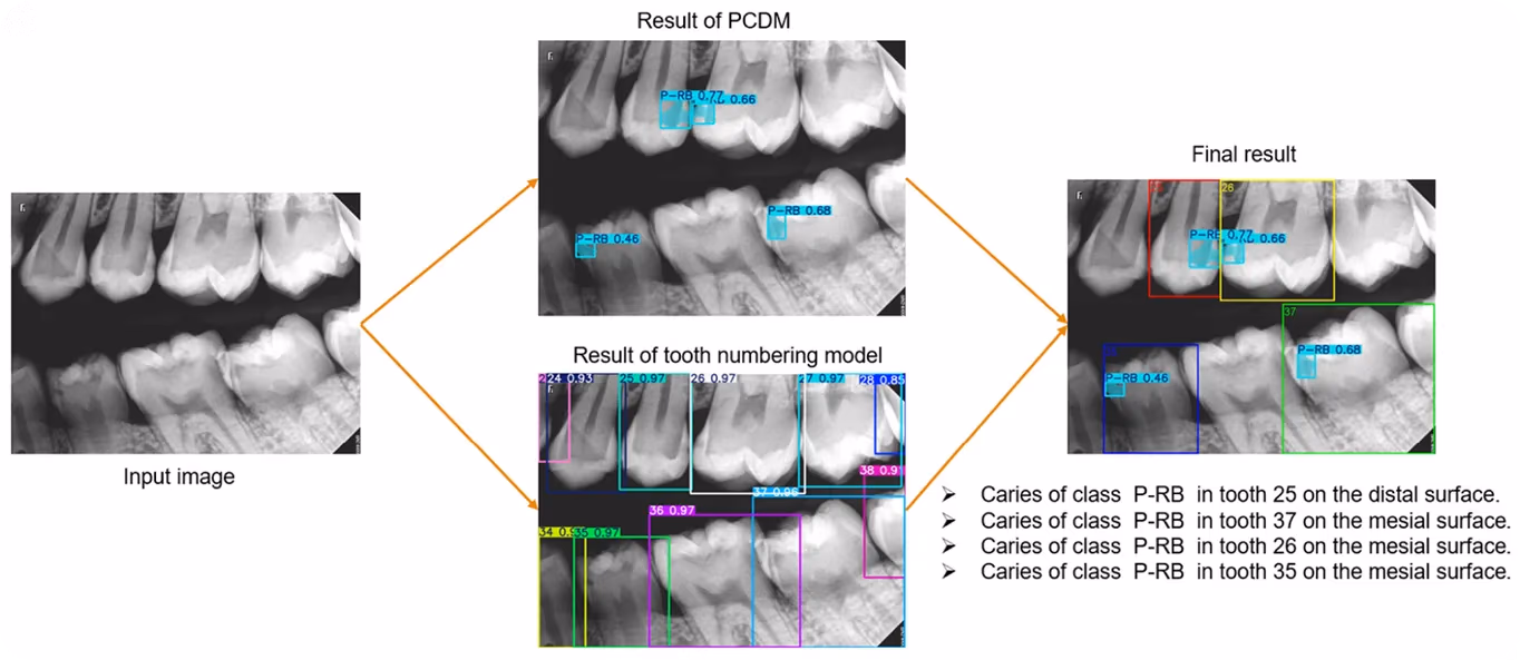
Fig 4. Detecting caries in dental X-rays using YOLO11 (Source)
Link to this sectionVision AI-driven robots in dental surgery#
Some dental procedures, like full-mouth reconstruction, can take several hours and leave both patients and dentists feeling tired. With the help of computer vision, dental robots are now being developed to assist with these procedures or even perform them independently with speed and accuracy.
For example, the world’s first fully automated dental procedure was completed by an AI dental robot in July 2024 in Colombia. The robot completed the full procedure in just 15 minutes. It used computer vision to create a detailed model of the patient’s mouth, identifying teeth, nerves, and cavities.
Based on this real-time data, the robot performed the surgery with a level of accuracy that would normally take several appointments. Although still in early stages, this technology shows the potential of vision AI to make dental care faster, more precise, and more comfortable in the future.
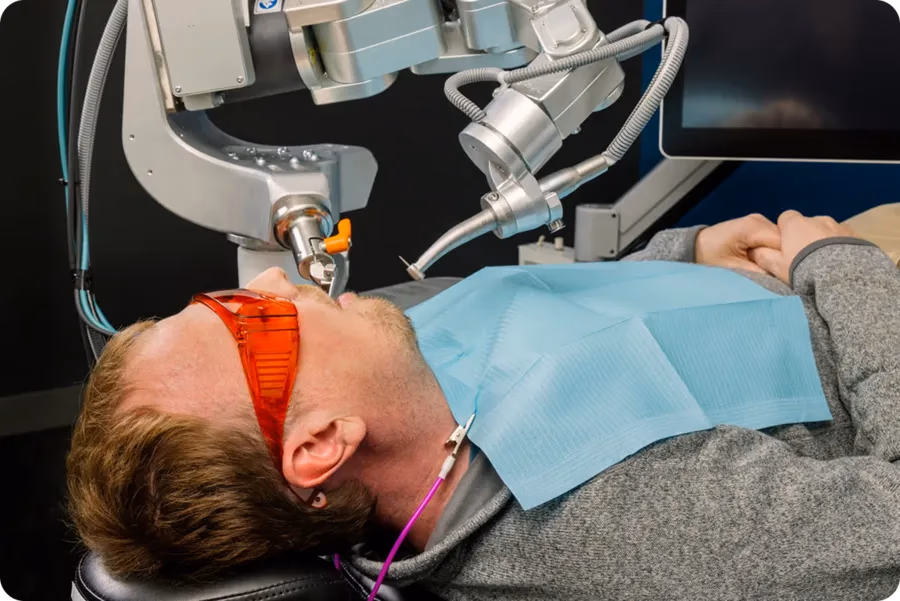
Fig 5. An autonomous robot performing a dental surgery (Source)
Link to this sectionFAQs about front tooth cavities#
Next, we’ll walk through some of the most common questions patients have about tooth decay in front teeth, along with the answers.
Link to this sectionWhy are front teeth prone to cavities?#
Front teeth are more prone to cavities because they have thinner enamel, and plaque often collects near the gumline. Frequent consumption of sugary or acidic foods also weakens enamel, making the teeth more vulnerable to decay.
Link to this sectionCan front tooth cavities heal in the early stages?#
Yes. In their earliest stage, cavities may appear as white spots, and at this point, enamel can sometimes repair itself. Using fluoride toothpaste, receiving professional fluoride treatments, and maintaining good oral hygiene all support enamel remineralization.
Link to this sectionAre veneers or tooth-colored fillings noticeable?#
Veneers and tooth-colored fillings are not usually noticeable because they blend with the natural tooth, restoring the front surface appearance while maintaining a healthy smile.
Link to this sectionWhat is the risk of cavities spreading to other parts of the tooth?#
If left untreated, cavities can progress deeper, from enamel into dentin and eventually the pulp, leading to pain and possible infection. Regular dental check-ups and X-rays help catch decay early and prevent further spread.
Link to this sectionHow can computer vision help detect front tooth cavities?#
Computer vision can analyze X-rays and images of the tooth surface to detect early signs of cavities. By assisting dentists in identifying decay at an early stage, AI tools help preserve natural tooth structure and improve long-term oral health.
Link to this sectionKey takeaways#
Tooth decay in front teeth is a common oral health issue, but it can be managed with the right care. Good oral hygiene and regular dental check-ups go a long way towards protecting natural teeth. In addition to this, AI and computer vision can be used by dentists to spot problems earlier, prevent serious damage, and support healthier smiles for the future.
Join our community and explore our GitHub repository to discover more about AI. If you're looking to start your own vision AI project, check out our licensing options. You can also see how AI in healthcare and vision AI in retail are making an impact by visiting our solutions pages.