Detecting buckle fractures of the wrist with computer vision
Learn what a buckle fracture of the wrist is and why it’s common in children. Explore how AI and computer vision help doctors detect and treat fractures accurately.

A wrist buckle fracture, also known as a torus fracture or distal radius buckle fracture, is a common wrist injury that occurs when the wrist bone bends and slightly bulges instead of breaking completely. This type of fracture is most common in children, especially around the wrist.
While painful, buckle fractures are generally stable and heal quickly with simple treatment, such as wearing a splint. Traditionally, doctors have relied on X-rays to diagnose these types of injuries, but subtle fractures can sometimes be missed.
Today, a cutting-edge technology referred to as computer vision is being explored to improve diagnosis. Computer vision is a branch of artificial intelligence (AI) that enables computers to see and interpret images, such as X-rays, in a manner similar to human vision. By assisting doctors in accurately detecting wrist buckle fractures, computer vision can also help reduce unnecessary treatments.
In this article, we’ll take a closer look at what a buckle fracture is, why it’s so common in children, how it’s treated, and how computer vision is shaping the future of diagnosis and recovery. Let’s get started!
Link to this sectionWhat causes a wrist buckle fracture#
Before we dive into how computer vision is improving patient care for fractures, let’s first get a better understanding of what a wrist buckle fracture is, how it happens, and why it’s so common in children.
Wrist buckle fractures are common among children under the age of 12. Adult bones are harder and more brittle, while children’s bones are softer and more flexible. Also, children’s bones have a thinner, more flexible cortex (the hard outer layer of the bone) and a thicker periosteum (the protective covering that helps the bone heal).
Because of this flexibility, when a child falls on an outstretched hand, the bone doesn’t usually snap like it might in an adult. Instead, one side of the cortex compresses and bulges outward, creating what doctors call a buckle.
Buckle fractures occur in the distal radius (the larger bone near the wrist) and sometimes the ulna (the smaller bone alongside it). Even simple accidents like falling off a cycle, tripping on the playground, or awkward landings in contact sports can put enough pressure on a child’s wrist to cause this type of fracture.
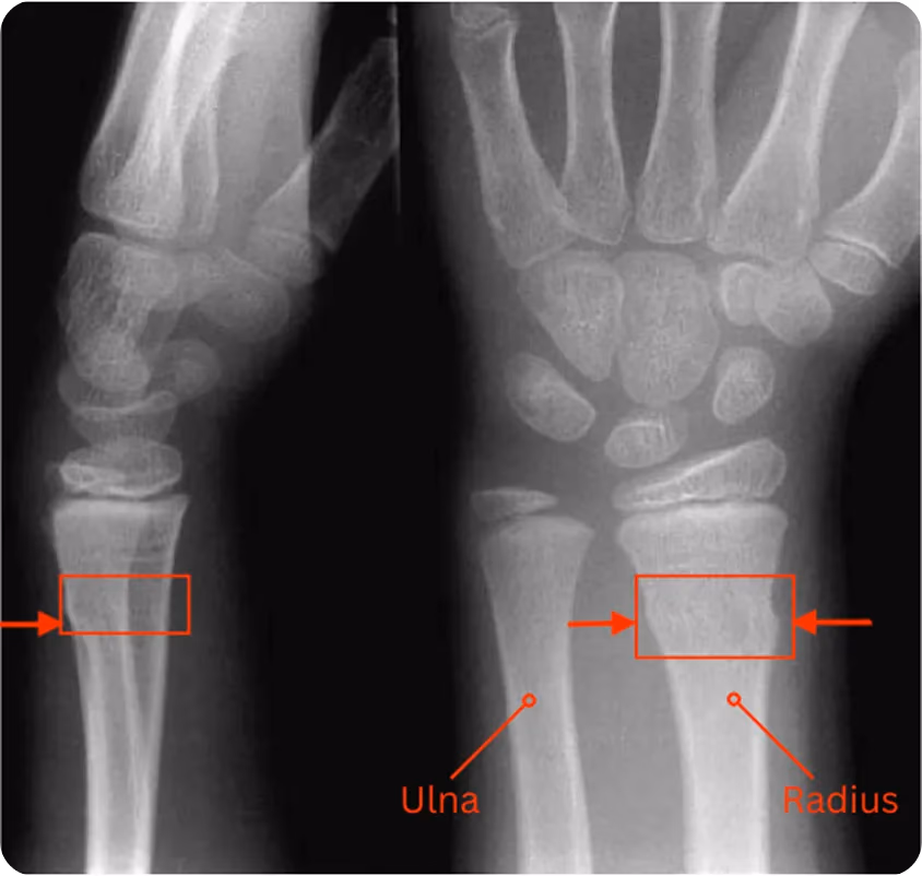
Fig 1. A buckle fracture of the wrist on the distal radius. (Source)
Unlike severe injuries, such as greenstick fractures or complete fractures, buckle fractures are incomplete fractures that typically heal quickly and rarely cause long-term problems. However, it’s still important to have the injury properly diagnosed and treated to ensure the bone heals correctly and to avoid complications.
Link to this sectionSpotting the signs of a wrist buckle fracture at home#
Now that we have an overview of what a wrist buckle fracture is, let’s look at how it’s usually treated before moving on to the challenges involved and the role of computer vision.
A buckle fracture can be hard to spot because the wrist doesn’t always look badly injured. The common signs are pain, swelling, and tenderness around the wrist. In some cases, there might be a small bump or slight change in shape, but often the wrist looks fairly normal.
What often confuses parents is that, even with a buckle fracture, a child may still be able to move their wrist fairly well. With a complete break, the wrist becomes unstable and movement is almost impossible, but a buckle fracture is more stable, so some motion is still possible.
This can make it easy to mistake a buckle fracture for a sprain. Both cause pain and swelling, but a fracture involves the bone, while a sprain affects the ligaments. If the pain doesn’t improve in a day or two, or keeps coming back when the child uses their wrist, it’s best to have it checked by a healthcare provider. Even though buckle fractures are less serious than other breaks, they still need proper care to heal well.
Link to this sectionTreatment: How doctors care for wrist buckle fractures#
Most buckle fractures heal quickly and don’t need ongoing treatment. Recovery mainly involves keeping the wrist stable and comfortable while the bone heals.
Doctors usually focus on three simple steps: splinting, pain relief, and a short follow-up to check progress. Let’s walk through these three steps.
Link to this sectionSplinting and immobilization#
Instead of a heavy plaster cast, orthopedic surgeons often use a removable splint that wraps around the wrist to keep it still. These wrist splints are easier for parents to manage, more comfortable for children, and are recommended by the NHS (the United Kingdom’s National Health Service). They can sometimes be removed for bathing or short breaks, depending on the doctor’s advice.
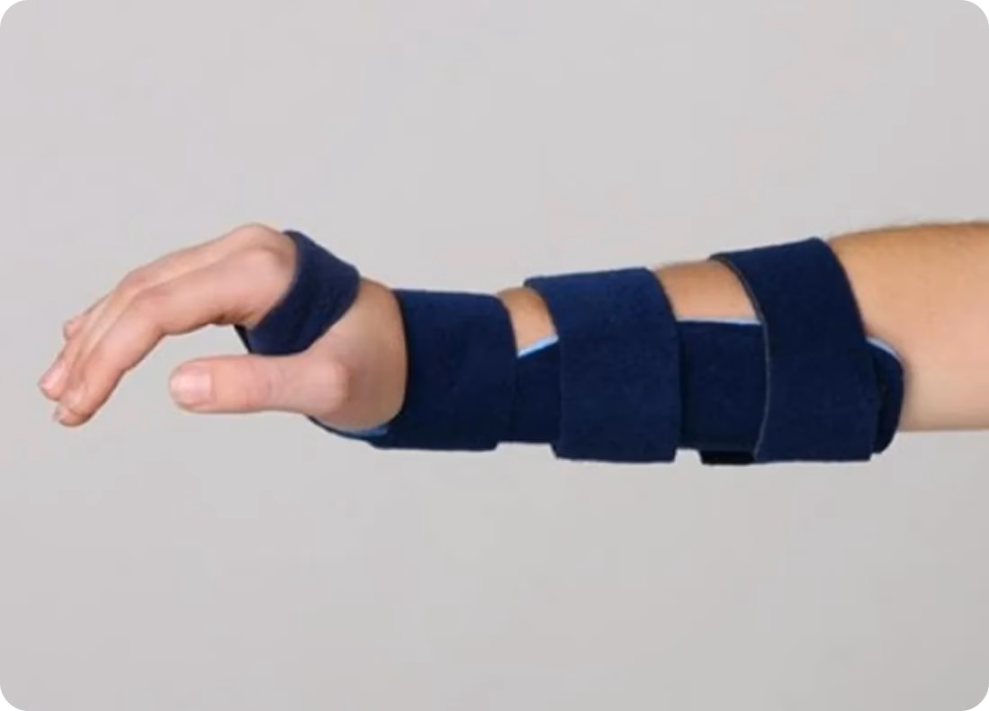
Fig 2. A removable wrist splint (Source)
Link to this sectionPain relief#
In the first few days, the fracture may be sore, but safe doses of painkillers such as ibuprofen or paracetamol are often enough. With the wrist supported in a splint and the pain managed, most children start feeling more comfortable quickly.
Link to this sectionRecovery and follow-up#
Buckle fractures usually heal within a few weeks and rarely cause lasting problems. Some children may need a follow-up appointment, where a doctor checks that the bone is healing properly.
Once the splint is off and the pain has eased, children can return to regular play, though contact sports like football or gymnastics may require a little extra time. In most cases, the fracture heals fully with no long-term effects.
Link to this sectionThe need for computer vision in healthcare#
In hospitals and clinics, doctors review hundreds of medical images every day, such as X-rays, CT scans, and MRIs. While these images reveal vital information, subtle details can be overlooked during manual review. Computer vision models are designed to interpret those images and other visual information with speed and precision.
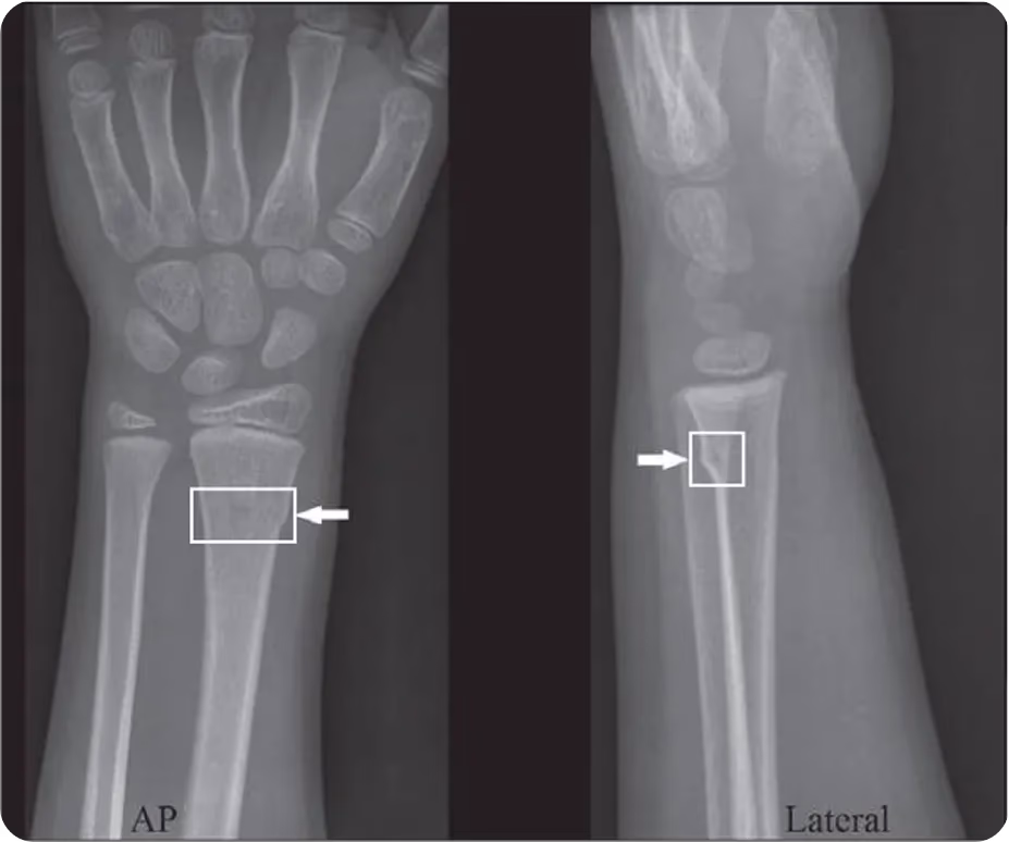
Fig 3. X-ray showing a buckle fracture of the wrist (Source)
For example, vision AI models like Ultralytics YOLO11 and Ultralytics YOLOv8 support tasks such as object detection (locating objects within an image), image classification (assigning a label to the whole image), and instance segmentation (outlining the exact shape and boundaries of objects). When trained on a custom set of X-ray images, YOLO11 and YOLOv8 can learn to spot the subtle signs of buckle fractures, making them more useful for diagnosis.
In the context of fractures, object detection can help pinpoint the exact location of the injury on an X-ray, while image classification can determine whether the scan is normal or shows a fracture, and can even identify the type of fracture. Instance segmentation can go a step further by outlining the precise shape and boundaries of the fracture, giving doctors a clearer picture of how much of the bone is affected.
Link to this sectionWhy use Ultralytics YOLO models for wrist fracture detection?#
You might be wondering why, with so many computer vision models available today, an Ultralytics YOLO model like YOLO11 should be used. YOLO models such as YOLO11 and YOLOv8 are popular because they combine speed, accuracy, efficiency, and precision in a way that makes them practical for real-world use. This is especially important in hospitals and clinics where doctors review large numbers of X-rays every day.
In fact, in a recent study focused on detecting pediatric wrist abnormalities, researchers compared several YOLO models (Ultralytics YOLOv5, YOLOv6, YOLOv7, and YOLOv8) with traditional two-stage object detection approaches like Faster R-CNN and even conventional CNN classifiers.
The results showed that the YOLO family of models not only worked faster but also picked up fractures with higher accuracy. Among them, YOLOv8m performed especially well, achieving a sensitivity of 92% and a mean average precision (mAP) of 95% for fracture detection.
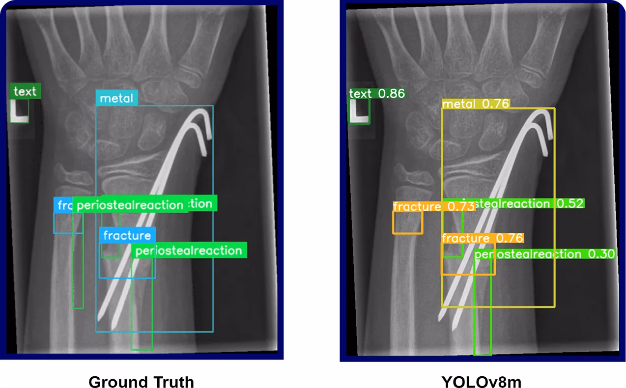
Fig 4. An example of using YOLOv8 to detect wrist fractures (Source)
Link to this sectionKey takeaways#
Buckle fractures are common in children but usually heal quickly with a splint and some rest. Doctors are now starting to use computer vision to spot these fractures more accurately, which means fewer missed diagnoses and quicker care. With the right treatment and a little help from AI, kids can get back to their normal activities with confidence.
Join our community and check our GitHub repository to learn more about AI and computer vision. Explore our solutions pages to learn more about computer vision in healthcare and AI in agriculture. Check out our licensing options and start building your own computer vision model.