Medical Image Analysis
Explore how AI transforms medical image analysis. Learn to detect anomalies and segment scans using Ultralytics YOLO26 for faster, more accurate diagnostics.
Medical Image Analysis is a specialized branch of computer vision (CV) and artificial intelligence (AI) focused on interpreting and extracting meaningful insights from medical scans. By leveraging advanced algorithms, this field automates the detection of biological structures and anomalies in complex imaging data, such as X-rays, Computed Tomography (CT), Magnetic Resonance Imaging (MRI), and ultrasound. The primary goal is to assist radiologists and clinicians by providing accurate, quantitative data to support diagnostic decisions, treatment planning, and long-term patient monitoring.
Link to this sectionCore Techniques and Methodologies#
The workflow typically begins with the ingestion of high-resolution images, often stored in the standardized DICOM format. To ensure algorithms perform optimally, raw scans usually undergo data preprocessing techniques like normalization and noise reduction. Modern analysis relies heavily on deep learning (DL) architectures, particularly Convolutional Neural Networks (CNNs) and Vision Transformers (ViT), to execute specific tasks:
- Object Detection: This involves locating specific features, such as identifying a nodule in a lung scan. The model predicts a bounding box around the region of interest, highlighting potential issues for physician review.
- Image Segmentation: A more granular approach where the model classifies every pixel. This is crucial for delineating precise boundaries, such as separating a tumor from healthy tissue or mapping the ventricles of the heart using architectures like U-Net.
- Image Classification: The system assigns a diagnostic label to an entire image, such as categorizing a retinal scan as either healthy or indicative of diabetic retinopathy.
Link to this sectionReal-World Applications in Healthcare#
Medical image analysis has moved from theoretical research to practical deployment in hospitals and clinics.
-
Oncology and Tumor Tracking: Advanced models like Ultralytics YOLO26 are employed to detect malignant growths in MRI or CT scans. For example, using the Brain Tumor Detection dataset, AI systems can identify lesions with high recall, ensuring subtle anomalies are not overlooked during routine screenings.
-
Surgical Robotics: During minimally invasive procedures, real-time pose estimation helps robotic systems track surgical instruments relative to patient anatomy. This improves safety by ensuring tools remain within safe operating zones, often powered by low-latency platforms like NVIDIA Holoscan for immediate feedback.
The following Python snippet demonstrates how to load a trained model and perform inference on a medical scan to identify anomalies:
from ultralytics import YOLO

# Load a custom YOLO26 model trained on medical data
model = YOLO("yolo26n-tumor.pt")

# Perform inference on a patient's MRI scan
results = model.predict("patient_mri_scan.jpg")

# Display the scan with bounding boxes around detected regions
results[0].show()
Link to this sectionChallenges and Considerations#
Applying AI to medicine presents unique hurdles compared to general imagery. Data privacy is a critical concern, requiring strict adherence to legal frameworks like HIPAA in the US or GDPR in Europe. Additionally, medical datasets often suffer from class imbalance, where examples of a specific disease are rare compared to healthy control cases.
To overcome data scarcity, researchers frequently use data augmentation to artificially expand training sets or generate synthetic data that mimics biological variability without compromising patient identity. Tools like the Ultralytics Platform facilitate the management of these datasets, offering secure environments for annotation and model training.
Link to this sectionDistinguishing Related Terms#
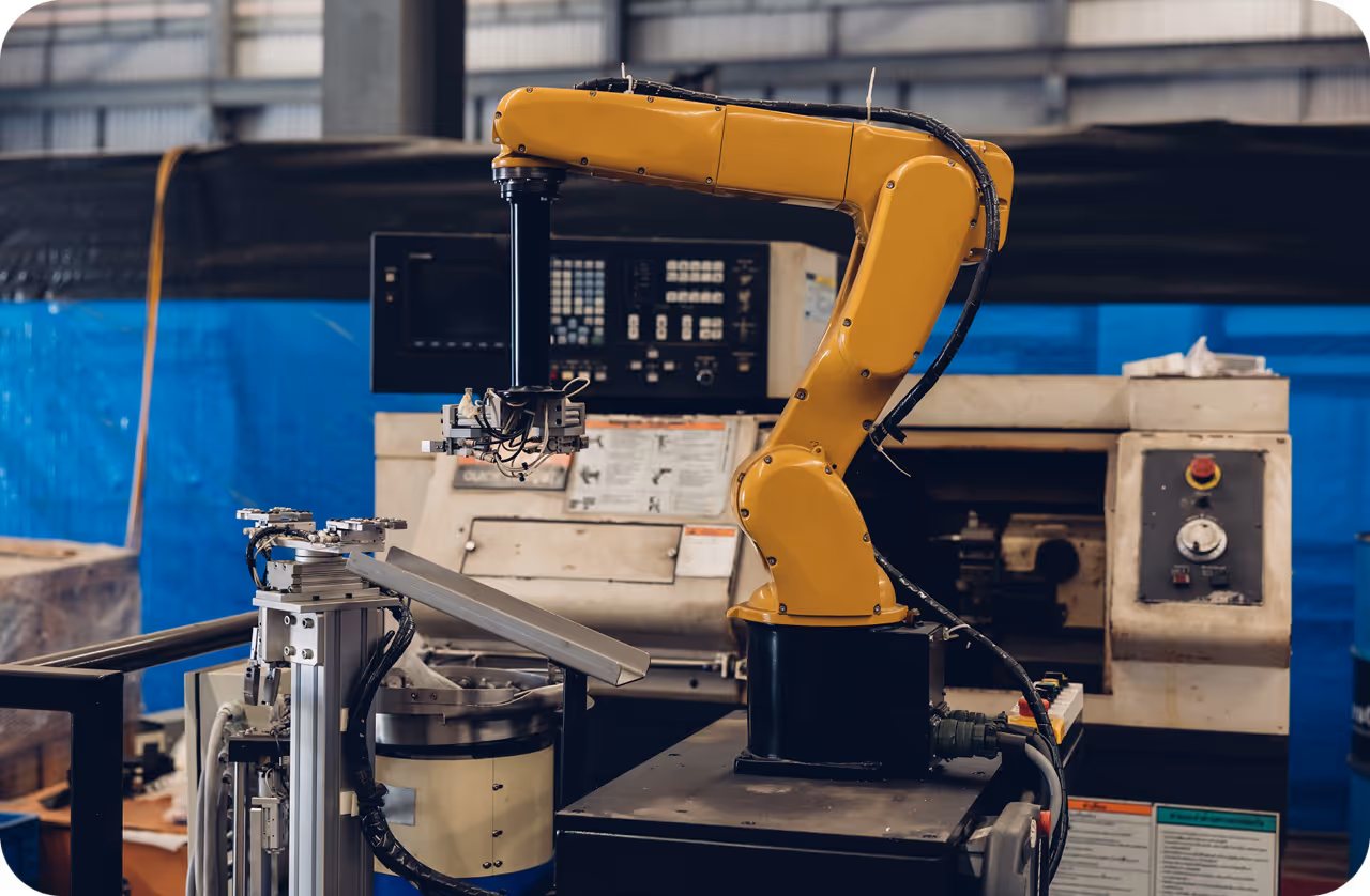
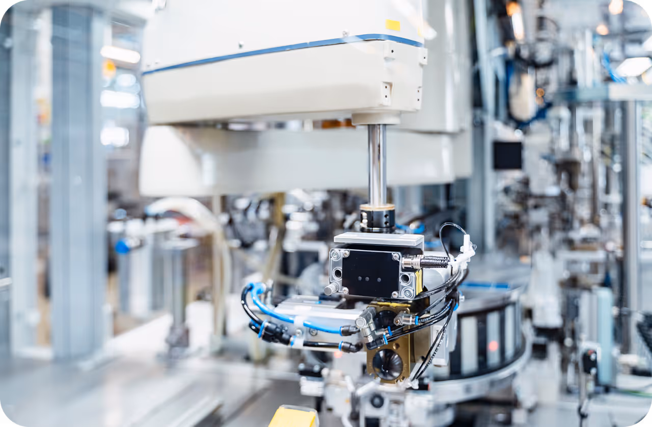
- vs. Machine Vision: While both involve analyzing images, machine vision typically refers to industrial applications, such as inspection on assembly lines. Medical image analysis deals with biological variation and requires probabilistic interpretation rather than pass/fail logic.
- vs. Biomedical Imaging: Biomedical imaging refers to the hardware and physics of creating the image (e.g., the MRI machine itself), whereas analysis focuses on the software algorithms that interpret the resulting data.
Regulatory bodies such as the FDA are increasingly establishing guidelines to ensure these AI in healthcare solutions are safe, effective, and free from algorithmic bias before they reach patient care.