Exploring applications of computer vision in microbiology
Learn how computer vision in microbiology can support accurate cell analysis, efficient colony counting, and improved diagnostics in research labs.

Observation is a key part of microbiology, where researchers analyze cells under microscopes, track bacterial colonies, and monitor microbial growth. These types of observational tasks are essential to both research and diagnostic processes.
Thanks to recent innovations in digital imaging and automation, laboratories are now producing more visual data than ever before. For instance, a high-resolution microscope can easily capture thousands of images for a single study. Each image contains minute and important details.
However, going through them individually can be a slow and inconsistent process. This increase in data has created a need for faster and more reliable image analysis.
One of the key technologies helping to automate this process is computer vision, which enables computers to interpret and analyze visual information from images or videos. In particular, vision AI models like Ultralytics YOLO11 are being used to support microbiology research by classifying cells, counting bacterial colonies, and tracking microbial growth.
In this article, we’ll explore how computer vision in microbiology is enhancing lab workflows and enabling scientists to work more efficiently and consistently. Let’s get started!
Link to this sectionThe role of computer vision in microbiology#
Computer vision tasks like object detection and image classification, powered by models such as YOLO11, can be used to detect patterns, highlight important features, and automate repetitive lab tasks that would otherwise take up valuable time and effort. Before we dive into specific applications, let’s take a closer look at how computer vision is being used in microbiology.
Link to this sectionCell classification using computer vision#
Cell classification is one of the most critical image-based tasks in microbiology. Labs often use stained images to help identify cell types, detect signs of infection, and highlight specific cell features under the microscope. Manual reviews take time and can be challenging to scale. Many labs are now using computer vision to detect, segment, and classify cells automatically to address this.
For example, at University Hospital Monklands in Scotland, a pilot program used computer vision to improve cervical cancer screening. Samples from patients who tested positive for Human Papillomavirus (HPV) were digitized and processed using vision AI models. The system analyzed cell structures and flagged any with unusual features for review by a medical expert.
This helped the team prioritize high-risk samples earlier in the workflow. As a result, slide reviews became faster and more focused, and they were able to handle more screenings without changing how samples were prepared or submitted.
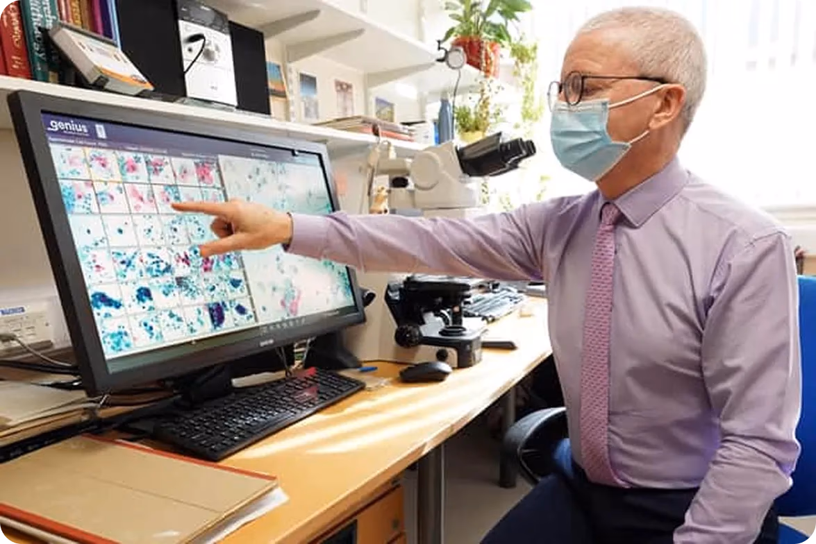
Fig 1. Cell classification using computer vision can enhance AI-assisted cervical screening.
Link to this sectionColony counting automation and growth analysis#
Colony counting is a laboratory technique used to measure microbial growth and evaluate how samples respond to treatment. It is widely used in vaccine development, clinical testing, and food safety. The counting process can be complex when done manually, especially when colonies overlap or plate volumes increase.
To streamline this, computer vision tasks like instance segmentation can be used to outline colony boundaries, measure their size, and count each colony based on its shape and spread, even in cases of overlap. This makes the review process faster and more consistent across batches.
For instance, an internationally recognized vaccine research facility is using ProtoCOL 3, an advanced colony counter powered by computer vision. The system scans multi-well plates and analyzes the colonies that survive after antibody exposure. With this automation, the facility increased its output from analyzing 16 plates to over 300 per day.
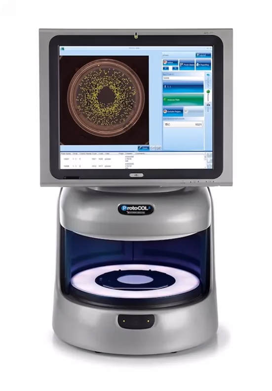
Fig 2. A look at ProtoCOL 3 - an example of colony counting automation (Source: labbulletin.com).
Link to this sectionMicroscopy image enhancement with AI#
Microbiologists regularly use microscopes to observe the structure and behavior of microbial cells. However, microscope images are often difficult to analyze due to overlapping cells, faint boundaries, and visual noise.
This is exactly why labs are turning to computer vision tools that enhance image clarity by applying techniques like image segmentation and noise reduction before processing them for tasks like colony counting or cell classification.
Beyond this, AI-driven image enhancement is being used to improve the clarity of low-resolution images of small cellular structures, such as mitochondria and brain tissue. This makes it possible for scientists to analyze important details in real time, speeding up research and improving diagnostic accuracy.
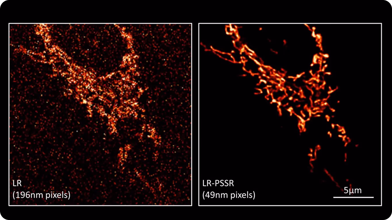
Fig 3. A mitochondrial network in a cancer cell, shown in low resolution (left) and enhanced by AI (right).
Link to this sectionReal-world applications of computer vision in microbiology#
Now that we've discussed how computer vision is used in microbiology, let's dive into some real-world applications.
Link to this sectionPharmaceutical research enabled by computer vision#
Any medication we take when we're not feeling well, even something as simple as a cold remedy, has a huge effort behind it. Pharmaceutical research is the process of discovering and developing new drugs to treat diseases, and a key part of this involves testing how compounds affect microbial cells. Scientists often grow bacteria on culture plates to see if a drug can stop microbial growth.
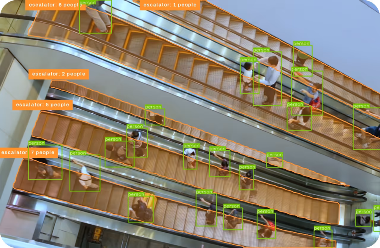
We are now seeing computer vision models like YOLO11 being used to accelerate the analysis of culture plates through object detection. YOLO11 can detect and count cells, and these insights can, in turn, be used to track their growth or shrinkage in response to treatments, making the research process faster and more efficient.
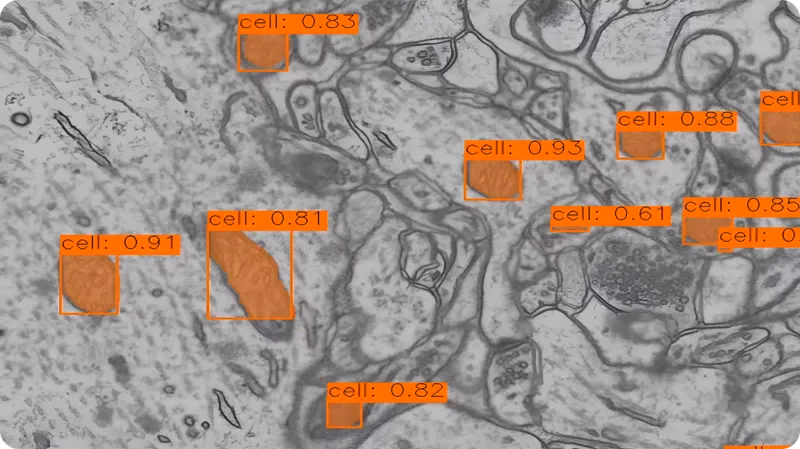
Fig 4. An example of using YOLO11 to detect cells.
Link to this sectionVision-driven clinical diagnostics#
While pharmaceutical research deals with discovering and testing new drugs, diagnostic labs focus on analyzing biological samples, such as blood, to detect signs of infection or disease. The purpose behind diagnostic labs is to provide accurate, timely information that helps in diagnosing conditions, monitoring disease progression, and guiding treatment decisions.
Though the core insights from these analyses may differ, the investigations themselves are quite similar, which is why computer vision is also impactful in this field. For example, in blood analysis, computer vision can be used to classify blood cells, such as red blood cells, white blood cells, and platelets, automatically.
By applying image classification and object detection, vision AI models can accurately detect and categorize these cells, streamlining the review process and helping researchers or clinicians focus on areas that need immediate attention.

Fig 5. Using computer vision to detect blood cells.
Link to this sectionPros and cons of computer vision in microbiology#
Computer vision enables microbiology labs to streamline image-based tasks, improving efficiency and consistency. It accelerates analysis, reduces manual labor, and enhances repeatability across processes. Here are some other key benefits of using computer vision in microbiology:
- Cost efficiency: Automating image analysis reduces the need for additional personnel, lowering labor costs while increasing productivity.
- Fewer manual errors: Visual errors and inconsistent observations are reduced, as models apply the same rules across every image.
- Supports remote and real-time use: Images can be processed and reviewed from different locations. This helps researchers to collaborate or monitor data in real time.
- Scalability: As data volumes increase, computer vision systems can easily scale to handle larger datasets without requiring proportional increases in labor or resources.
Despite these advantages, there are also a few limitations to consider. To make the most of vision AI tools, proper planning, support, and setup are vital. Here are a few key challenges to keep in mind:
- Initial cost and setup: Getting started with AI tools requires significant investment in hardware, software, and training, which can be a barrier for some labs.
- Data privacy and security: Handling sensitive data, especially in healthcare or clinical research, requires robust security measures to ensure compliance with privacy regulations.
- Integration with existing systems: Implementing AI solutions can be challenging if the new tools need to integrate with existing lab management systems or workflows.
- Ongoing maintenance and updates: AI models require continuous monitoring, updates, and fine-tuning to remain effective, which can be resource-intensive.
Link to this sectionThe road ahead for Vision AI in microbiology#
Computer vision in microbiology is moving toward tools that are easier to train and more practical to use in real lab settings. Researchers are focusing on models that need less data to get started and can adapt more quickly when lab conditions change.
One particularly fascinating area of progress is mobile microscopy. AI models are now being integrated into small devices that work outside of traditional lab setups. These systems capture microscope images and analyze them on-site, making them ideal for use in remote areas with limited infrastructure.
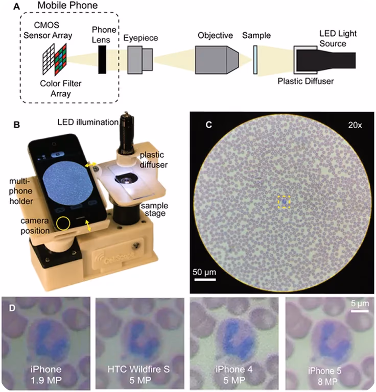
Fig 6. Using a smartphone-based microscope for cell imaging (Source: journals.plos.org).
Link to this sectionKey takeaways#
As digital imaging becomes central to microbiological research, the demand for faster and more consistent analysis continues to grow. Computer vision helps meet this need by efficiently handling core tasks like cell classification, colony counting, and segmentation with speed and accuracy.
Many labs have already transitioned from manual reviews to AI-supported systems. For labs dealing with high sample volumes or tight timelines, computer vision is quickly becoming a practical solution. These tools are easy to integrate into existing workflows, allowing labs to adopt them without major changes.
Join our growing community! Explore our GitHub repository to dive deeper into AI. If you are interested in leveraging computer vision, check out our licensing options. Learn about computer vision in healthcare and AI in manufacturing on our solutions pages!